Introduction
Key Takeaways
- Ultrasound is a safe, painless, and non-invasive imaging technique.
- It uses sound waves, not radiation, making it safe for pregnant women and children.
- Proper preparation, such as fasting or drinking water, is essential for clear results depending on the type of scan.
If your doctor has recommended an ultrasound scan, you might have questions about what the procedure entails, how to prepare, and what the results will show. Ultrasounds are one of the most common and versatile diagnostic tools used in modern medicine, providing real-time images of the inside of your body without the use of radiation.
At De Rock Hospital & Diagnostic Center, our advanced Ultrasound department is dedicated to providing clear, accurate imaging in a comfortable and reassuring environment. Here is everything you need to know before your scan.
What Is an Ultrasound Scan?
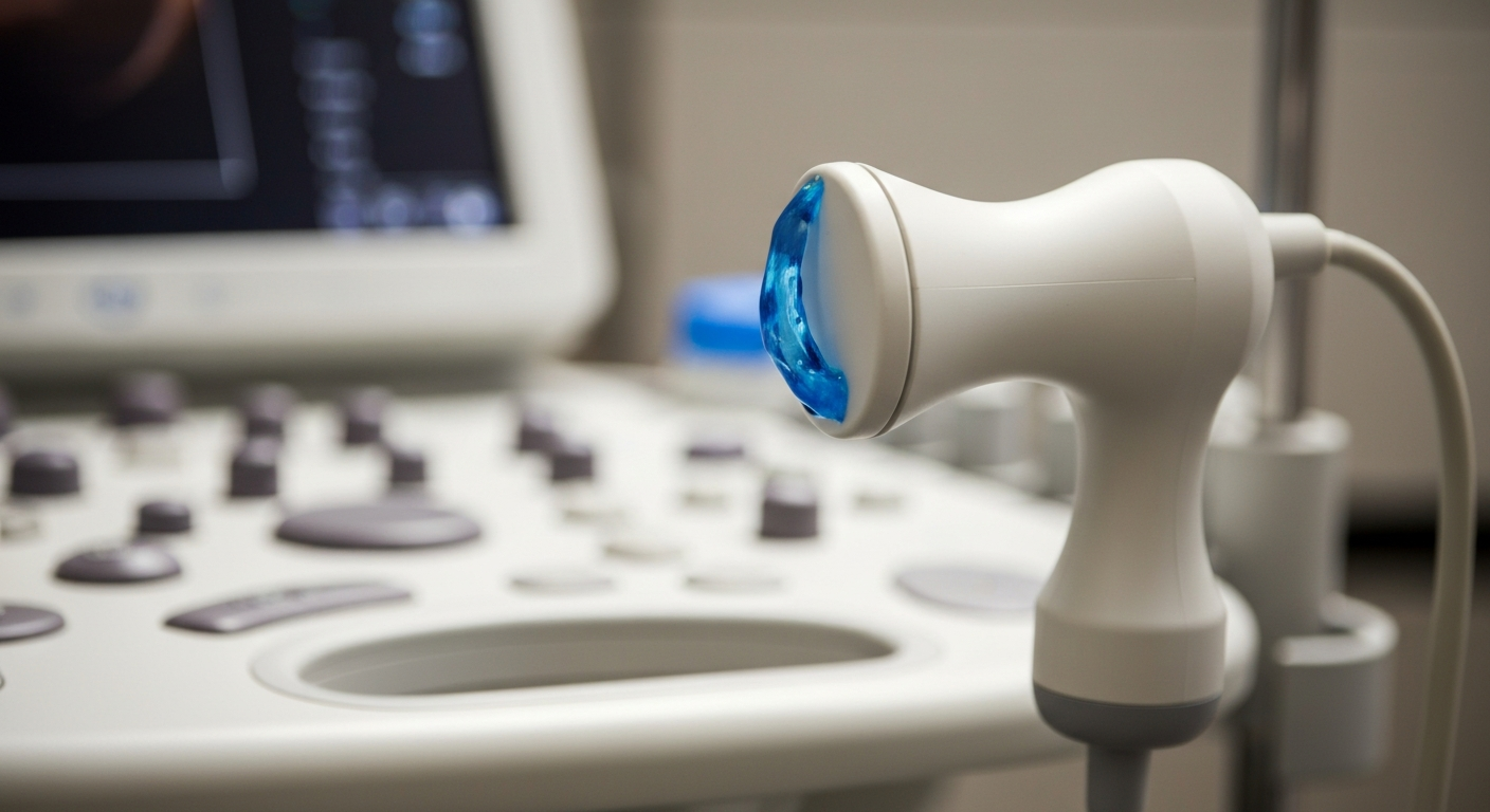
An ultrasound scan, also known as sonography, uses high-frequency sound waves to capture live images from the inside of your body. A small device called a transducer is placed against the skin. It emits sound waves that bounce off your internal organs and tissues, creating echoes that a computer translates into detailed images on a screen.
Why It Matters
Ultrasounds are crucial for diagnosing a wide variety of conditions quickly and safely. Because they capture images in real-time, they can show the structure and movement of the body's internal organs, as well as blood flowing through blood vessels. This allows doctors to identify abnormalities, monitor fetal development, and guide minimally invasive procedures.
Common Types of Ultrasound Scans
Ultrasounds can be used to examine many different parts of the body. Common types include:
- Abdominal Ultrasound: Examines organs such as the liver, gallbladder, kidneys, pancreas, and spleen.
- Pelvic Ultrasound: Evaluates the uterus, ovaries, prostate gland, and bladder.
- Obstetric Ultrasound: Monitors the health and development of a fetus during pregnancy.
- Doppler Ultrasound: Assesses blood flow through arteries and veins to detect blockages or clots.
- Thyroid Ultrasound: Evaluates the thyroid gland in the neck for nodules or abnormalities.
How to Prepare for Your Scan
Preparation varies significantly depending on the part of the body being examined. Your doctor or our diagnostic team will provide specific instructions, but general guidelines include:
- For an Abdominal Ultrasound: You may be asked to fast (eat or drink nothing) for 8 to 12 hours before the test. This prevents undigested food and gas from blocking the sound waves.
- For a Pelvic or Obstetric Ultrasound: You will likely need to drink several glasses of water before the exam and avoid urinating. A full bladder pushes the intestines out of the way, providing a clearer view of the pelvic organs.
- For other scans (like Thyroid or Vascular): Usually, no special preparation is required.
Always wear loose, comfortable clothing. You may be asked to change into a hospital gown for the procedure.
What to Expect During the Procedure
An ultrasound is a straightforward and generally painless procedure that typically takes between 15 and 45 minutes.
- You will lie down on an examination table.
- A sonographer will apply a clear, water-based gel to the area being examined. The gel helps the transducer make secure contact with the body and eliminates air pockets.
- The sonographer will press the transducer firmly against your skin and move it back and forth over the area of interest.
- You may feel mild pressure, but you should not feel pain. If a full bladder is required, you may feel some discomfort, but you will be able to empty it immediately after the scan.
- Once the imaging is complete, the gel is wiped off, and you can resume your normal activities immediately.
Understanding Your Results
After the scan, a specialist doctor (a radiologist) will analyze the images and send a detailed report to your referring physician. Your doctor will then discuss the findings with you, explain what they mean for your health, and recommend any necessary follow-up steps or treatments.
Conclusion
An ultrasound scan is a powerful, safe, and routine diagnostic tool that provides invaluable information about your health. By understanding the process and following preparation instructions, you can ensure the clearest possible images. At De Rock Hospital & Diagnostic Center, our expert sonography team is here to guide you through the process with professionalism and care.
Medical Disclaimer
This article is for informational purposes only and is not a substitute for professional medical advice, diagnosis, or treatment. Always seek the advice of your physician or other qualified health provider with any questions you may have regarding a medical condition.